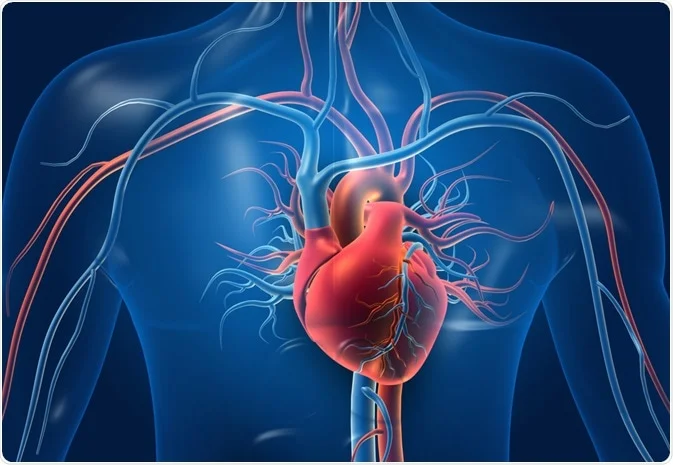
Nuclear stress testing is a diagnostic tool that provides detailed insight into how well the heart functions under physical stress. By using a small amount of radioactive material and imaging technology, doctors can evaluate blood flow, detect blockages, and assess overall heart health. Understanding the role of cardiac blood flow testing highlights its significance in identifying heart conditions early and guiding effective treatment plans.
What Is a Nuclear Stress Test?
Nuclear stress testing uses a small amount of radioactive material to create detailed pictures of your heart. The procedure combines physical exercise with nuclear imaging to show how blood flows to your heart muscle during rest and activity. During the test, doctors inject a safe radioactive tracer into your bloodstream. This tracer helps create clear images of your heart using a special camera.
The test shows which areas of your heart receive adequate blood flow and which areas may have blocked or narrowed arteries. Cardiac blood flow testing is more detailed than regular stress testing because it provides visual images of blood flow patterns. The radioactive material used is completely safe and leaves your body naturally within a few days.
When Do Doctors Recommend It?
Doctors recommend nuclear stress testing when patients have symptoms that suggest heart problems. This test helps identify issues that only appear when the heart works harder during exercise or stress. Common reasons for recommending stress testing include chest pain that occurs with activity and shortness of breath during exercise. Another key reason may be abnormal results on other heart tests. Your cardiologist may also suggest this test if you have risk factors for heart disease, such as diabetes, high blood pressure, or a family history of heart problems.
What Should You Expect?
Your cardiologist will provide specific instructions about eating, drinking, and taking medications before your appointment. On the day of your test, a technician will place small electrode patches on your chest to monitor your heart rhythm. You will then receive an injection of the radioactive tracer through a small tube in your arm. After waiting about an hour for the tracer to circulate, you will lie on a table while a special camera takes pictures of your heart at rest.
Next comes the stress portion of the test. You will walk on a treadmill that gradually increases in speed and incline until your heart rate reaches the target. At peak exercise, you receive another injection of the radioactive tracer. About an hour after exercise, the camera takes more pictures to compare blood flow during rest and stress.
How Does It Help Me?
The results from the test provide doctors with detailed information about your heart’s blood supply and function. The images clearly show areas where blood flow is normal, reduced, or blocked completely. Normal results indicate that blood flows well to all parts of your heart during both rest and exercise. This means your coronary arteries are not significantly blocked and your heart muscle is healthy.
Abnormal results may show areas of your heart that receive less blood during exercise compared to rest. This pattern often indicates narrowed or blocked coronary arteries that limit blood flow when your heart needs it most. The test can also identify areas of damaged heart muscle from previous heart attacks.
Schedule Your Nuclear Stress Testing Today
Cardiac blood flow testing is a fundamental tool for diagnosing heart conditions that may not be detected with other tests. The procedure is safe, accurate, and provides key information that helps doctors develop effective treatment plans. If your doctor has recommended nuclear stress testing or if you have concerns about your heart health, contact a trusted cardiology practice near you today to schedule your appointment with a specialist.
- Must-Have Candle Making Supplies for Seasonal Scents
- Affordable Paths to Homeownership
- Gamerxo Dot Com: The Complete Guide to a Trusted Gaming Platform for U.S. Gamers
- Husziaromntixretos: A Complete and In-Depth Guide to Understanding This Emerging Digital Concept
- The Benefits of Seeing a Dermatologist for Children


